
Figure 8.
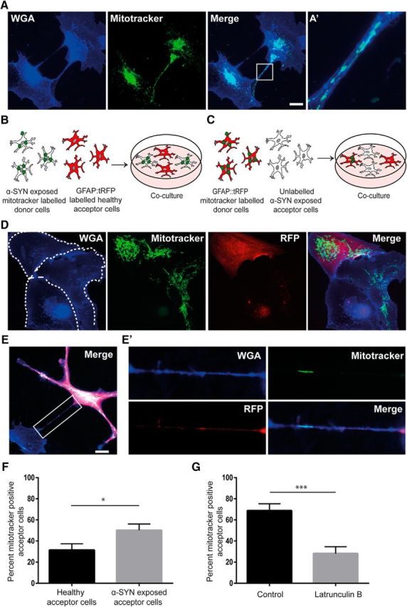
Healthy astrocytes deliver mitochondria to α-SYN oligomer-exposed astrocytes. Using Mitotracker transfection, we could demonstrate that mitochondria were present within TNTs connecting the astrocytes (A). A close-up of the white rectangle is shown in A′. Coculture experiments were performed using two different set-ups. Either, GFAP::tRFP astrocytes (red acceptor cells) were cocultured with Mitotracker-transfected, α-SYN oligomer-exposed astrocytes (donor cells) (B) or α-SYN oligomer-exposed astrocytes (acceptor cells) were cocultured with GFAP::tRFP, Mitotracker-transfected astrocytes (red donor cells) (C). Using immunocytochemistry, we identified transfer of mitochondria between the GFAP::tRFP astrocytes and the unlabeled astrocytes via direct contact (dashed areas show the outline of separate cells) (D) and TNTs (E), a close-up of the white rectangle is shown in E′. Quantification of the number of Mitotracker+ acceptor cells in the two coculture setups indicated that the healthy astrocytes transferred significantly more mitochondria to α-SYN oligomer-exposed cells than the other way around (F). Addition of latrunculin B to the cocultures significantly decreased the percentage of Mitotracker+ acceptor cells (G). In addition, basal level of mitochondrial transfer was investigated in untreated control cultures. Compared with α-SYN oligomer-exposed cultures, the number of Mitotracker+ acceptor cells was significantly lower in the control cultures (see Fig. 8-1). Scale bars: A, 20 μm; D, E, 20 μm. Data are presented as mean ± SEM from three independent experiments and the levels of significance were set to *p < 0.05, **p < 0.01, and ***p < 0.001 (F, G).