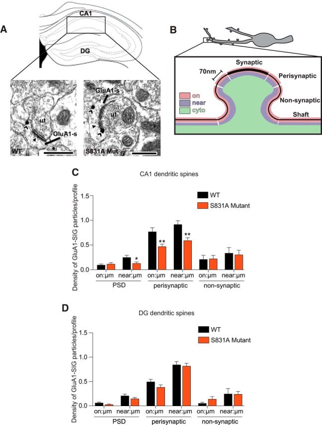
Figure 5.

S831A GluA1 mutant mice have lower levels of GluA1 near the PSD and on and near the plasma membrane of perisynaptic sites of dendritic spines in the hippocampal CA1 subregion after impaired cocaine CPP extinction. A, Representative electron micrographs from stratum radiatum of CA1 of WT and S831A mutant mice after cocaine CPP extinction showing GluA1 SIG particle labeling in spines (sp) contacted by unlabeled axon terminals (ut). Scale bars, 500nm. B, Schematic figure demonstrating the criteria used to apportion SIG particles when quantifying GluA1 localization in dendritic spines by electron microscopy. C, In dendritic spines, significantly lower densities of GluA1 SIG particles were seen near, but not on the plasma membrane of the PSD in S831A mice compared with WT mice. Moreover, significantly lower densities of GluA1 SIG particles were observed on and near the plasma membrane of perisynaptic sites in S831A mice compared with WT mice. No difference in density of GluA1 SIG particles was observed on or near the plasma membrane of nonsynaptic sites. D, No difference in densities of GluA1 SIG particles were observed at synaptic or perisynaptic sites of the dentate gyrus of S831A mice and WT mice *p < 0.05, **p < 0.01. Data are presented as mean ± SEM.
