Figure 6.
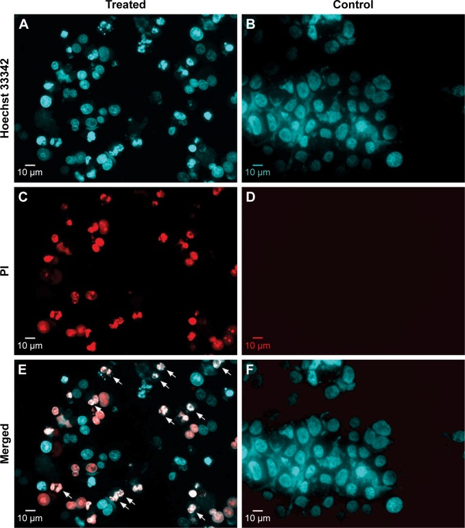
Disintegrated nuclei of H. odorata extract-treated cells.
Notes: After treatment with H. odorata extract (250 µg/mL) for 48 hours, HepG2 cells were harvested and stained with Hoechst 33342 (A and B) and PI (C and D) at a final concentration of 1 µg/mL for each dye. Nuclei of treated cells and control cells were then observed under a fluorescent microscope. Disintegrated nuclei are indicated with white arrows (E and F).
Abbreviations: H. odorata, Hopea odorata; PI, propidium iodide.

