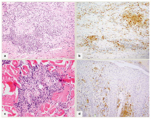
Fig 2.
Fig 2A. 200x magnification; granulomatous mycosis fungoides with prominent granuloma annulare-like appearance (palisading of histiocytes within papillary dermis), associated with atypical lymphocytes B. 200x magnification; an immunohistochemical stain for CD68 highlights the 25% component of histiocytes in this case C. 200x magnification; in syringotropic mycosis fungoides, a dense lymphoid infiltrate surrounds the eccrine glands and extends into glandular epithelium D. CD30 immunohistochemical stain, 200x; CD30+ mycosis fungoides with two distinct populations of lymphocytes characterized by CD30 positivity and small (intraepidermal) or large(dermal) cell size.

