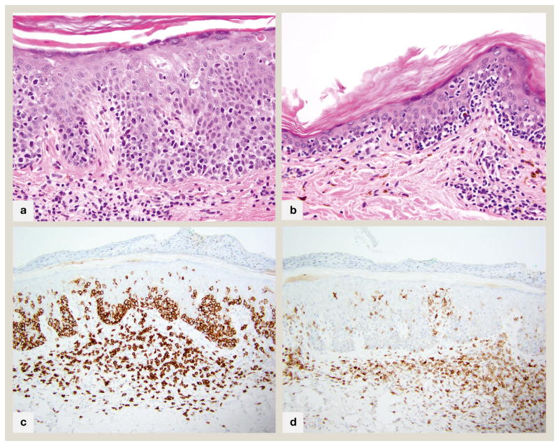
Fig 5.
CD8+ mycosis fungoides A. 400x magnification of an acanthotic lesion with prominent basal layer tagging and marked extension of lymphocytes into mid to upper layers of epidermis. A few necrotic keratinocytes are present B. 400x magnification of an atrophic lesion with marked basilar epidermotropism. Both A and B show few remaining pigmented basilar keratinocytes, but many superficial dermal melanophages, connoting basal layer destruction and melanin dropout. C. 200x CD8 immunohistochemical stain of case in part A showing diffuse staining of all epidermotropic lymphocytes D. 200x CD7 immunohistochemical stain of case in part A shows characteristic complete absence of this pan T-cell antigen

