Fig. 3.
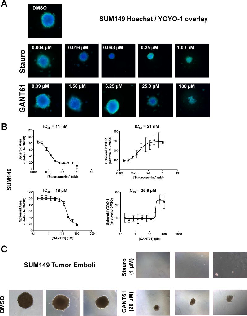
GANT61 disrupts formation of SUM149 tumor spheroids and tumor emboli. For the tumor spheroid assay, cells were seeded in ultralow attachment 384-well plates and treated 24 h later with either vehicle control (DMSO), staurosporine, or GANT61 at the indicated concentrations. Cells were grown for a further 72 h, spheroids stained with Hoechst/YOYO-1 and high content 3D images obtained using the CellInsight HCS platform. (A) representative images are shown for each treatment. Three independent experiments were carried out. (B) Data represent mean ± SD percent normalized to DMSO vehicle control for spheroid area (Hoechst) and spheroid YOYO-1 comprising a minimum of five replicate wells. Dose response curves were generated using non-linear regression and IC50 values determined in GraphPad Prism 6. (C) Tumor emboli: Representative images (10 × – scale bar = 100 microns) of SUM149 cells grown under tumor emboli conditions. Cells were treated at the time of plating with vehicle, staurosporine or GANT61 and cells grown for 7d. Images from at least two independent experiments comprising a minimum of three replicate wells.

