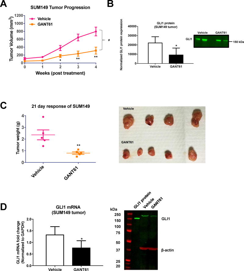
Fig. 5.
GANT61 treatment inhibits TN-IBC SUM149-derived xenograft tumor growth. (A) Nude NU/J mic were injected orthotopically with SUM149 cells into the mammary fat pad. Once tumors reached 5 × 5 mm (6 weeks) they were treated with vehicle or GANT61 (50 mg/kg i.p. 3× per week for n = five animals). Tumor response was assessed by weekly caliper measurements. Tumor volumes (Mean ± SEM) are shown for each time point. Statistical significance relative to respective control *P<0.05, **P<0.005, ***P<0.001 (Student t-test). Comparisons in tumor growth were also made by linear regression in GraphPad Prism 6.0. Statistical significance relative to control #P<0.05. (B) Quantification of the study shown in (A) for GLI1 protein expression by immunoblot analysis. Mean band intensity normalized to total protein (± SD) for vehicle compared to GANT61 treated tumor lysates (left panel). *P<0.05 (Student t-test). Representative immunoblot for GLI1 protein expression from three independent analyses of tumor cell lysates (B, right panel). (C) NOD SCID mice were injected orthotopically with SUM149 cells into the mammary fat pad. Once tumors were palpable, mice were treated with vehicle or GANT61 in vehicle (50 mg/kg i.p. once per day for n = five animals). After 21 days, tumors were excised and weighed. Weights are shown as Mean ± SEM. *P<0.05, **P<0.005, ***P<0.001 (Student t-test). (D) Quantification from study shown in (C) of GLI1 mRNA in the SUM149-derived tumors (left panel). *P<0.05 (Student t-test). Representative immunoblot from three independent experiments of tumor tissue lysates for GLI1 protein expression (D, right panel). Recombinant human GLI1 protein (GLI1 protein) was included as a positive control for the Western.

