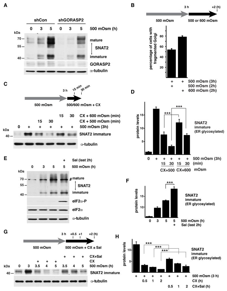
Fig. 4. Golgi apparatus fragmentation delays SNAT2 maturation in corneal epithelial cells.
A. Western blot analysis of extracts from cells expressing shRNA against GORASP2 exposed to hyperosmolar stress. Positions of protein size markers are indicated. B. Quantification of Golgi fragmentation in cells treated with 500 mOsm media for 3h and then switched to 500 or 600 mOsm media for additional 2h. C. Western blot analysis indicating the immature form of SNAT2 from cells treated with 500 mOsm media for 3h and then switched to 500 or 600 mOsm media in the presence of CX for the indicated times. D. Quantification of the data from panel C. SNAT2 signal intensities were normalized to α-tubulin E. Western blot analysis of extracts from cells cultured in 500 mOsm media for 5h with Sal003 (30 μM) added for the last2 h. A nonspecific band is indicated (#). F. Quantification of the immature SNAT2 levels from cells treated as in E as described above. SNAT2 signal intensities were normalized to α-tubulin. G. Western blot analysis showing the immature form of SNAT2 from cells treated with 500 mOsm media for 3h. The cells were then incubated with CX and Sal003 for the indicated times. H. Quantification of immature SNAT2 levels from cells treated as in G as described above. SNAT2 signal intensities were normalized to α-tubulin. Data in panels B, D, F, and H are represented as mean of 3 experiments ± SD.

