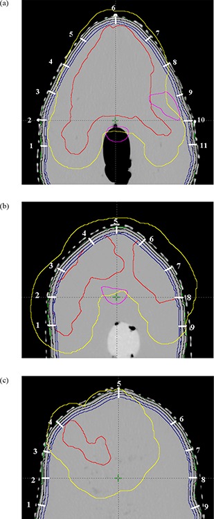
Figure 2.

The organs contoured on the CT slice images at isocentre, 5 cm inferior and superior to isocenter, with registration points for TLD placements: spinal cord (magenta color), the PTV to be delivered with 66 Gy (red). The magenta color contour to the right side of the CT axial slice represents the contralateral parotid (left parotid) to be saved; dark blue contours represent three strips of 2 mm thickness at three different depths of 2 mm, 4 mm and 6 mm from the skin of the phantom; yellow contour represents the region of interest which is defined by 1.4 cm extra margin from PTV for the defining of points of high‐ and low‐dose buildup regions.
