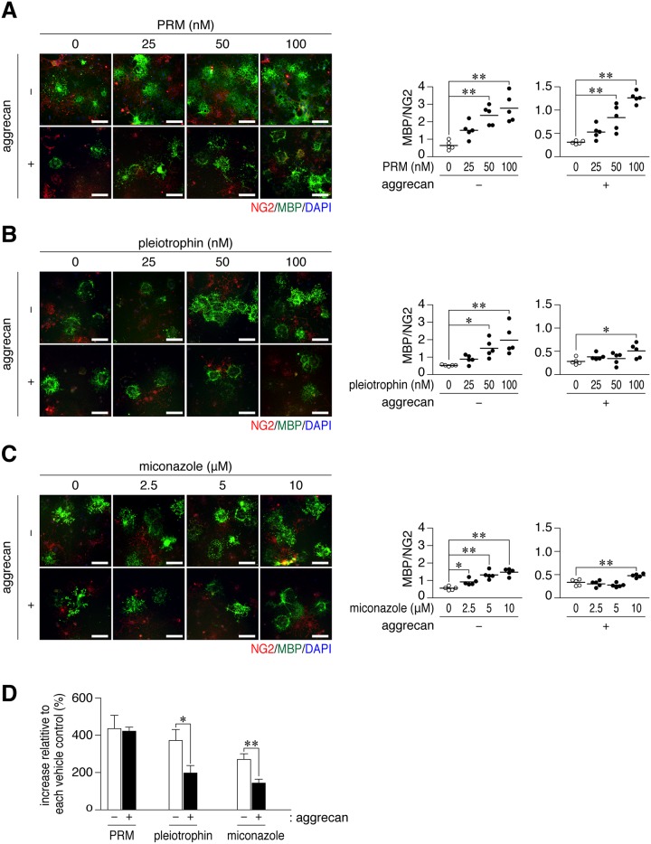
Fig 3. Protamine enhanced the differentiation of OL1 cells on aggrecan-coated dishes.
(A-C) Anti-NG2 and anti-MBP staining of OL1 cells. OL1 cells were cultured for 10 days on dishes coated with 50 μg/ml poly-L-ornithine (-), or a combination of 50 μg/ml poly-L-ornithine and 50 μg/ml aggrecan (+). Protamine (PRM, A), pleiotrophin (B), or miconazole (C) was added to differentiation medium at the indicated concentrations. Scale bars, 100 μm. The plots on the right side show the ratio of MBP-positive cells to NG2-positive cells in five independent cell cultures. *, p < 0.05 and **, p < 0.01, significant difference between the indicated groups (analysis of variance with Bonferroni’s post-hoc tests). (D) The percentage of the ratio of MBP to NG2 in 100 nM PRM, 100 nM pleiotrophin, or 10 μM miconazole to each vehicle is shown, demonstrating a small effect with pleiotrophin and miconazole on aggrean-coated substrates. Data were the mean with S.E. (error bars) from five independent experiments. *, p < 0.05 and **, p < 0.01, significant difference between the indicated groups (Student’s t-tests).

