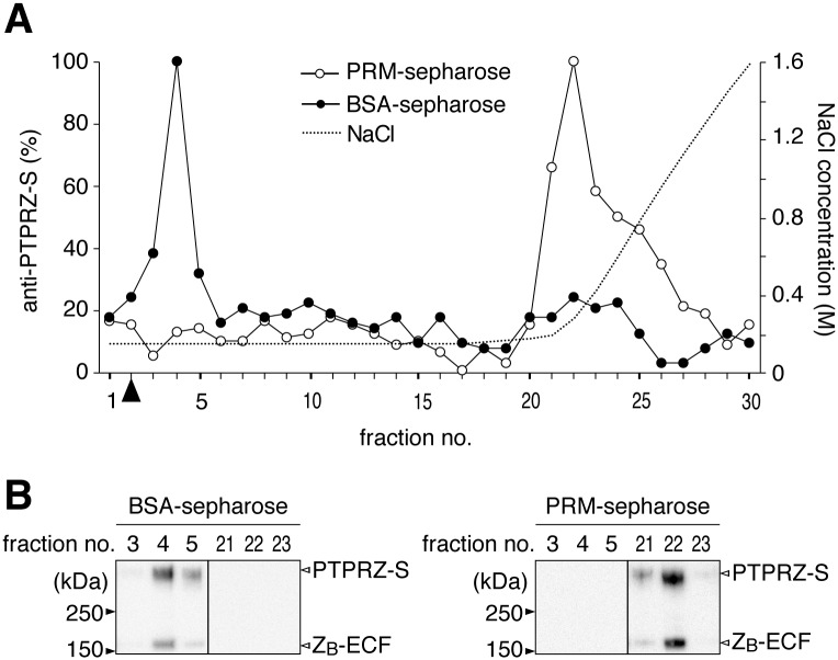
Fig 4. Extracellular portions of PTPRZ bearing CS were efficiently captured on PRM-immobilized resins.
(A) Elution profiles. Phosphate buffer extracts of mouse brains were applied to a Sepharose column immobilized with PRM or control BSA, and separated using 0.15 to 2.0 M NaCl gradient elution in 10 mM phosphate buffer, pH 7.3. Aliquots of the separated fractions were coated on microtiter wells, and the content of PTPRZ proteins was assessed using anti-PTPRZ-S. Arrowhead, the void volume. (B) Western blotting of eluted fractions. Relevant fractions separated by BSA-sepharose (left) and PRM-sepharose (right) were analyzed by Western blotting using anti-PTPRZ-S (a rabbit polyclonal antibody that recognizes an extracellular epitope of PTPRZ). Full-length blots are presented in S8 Fig.

