
Fig 3. The frequency of OCT4 expression in BM-MSCs decreases after co-culture with RECs as assayed by flow cytometry.
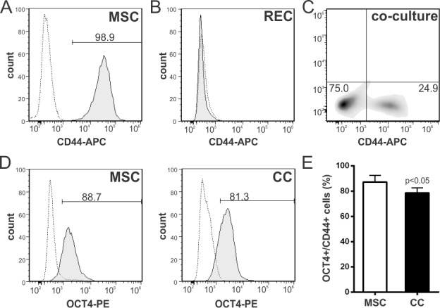
(A) BM-MSCs were stained with an APC-conjugated anti-mouse CD44 antibody. More than 98% of the cells are positive for this marker. Gray dotted line, isotype control; black line, antigen staining. (B, C) The CD44 antibody does not cross react with RECs and therefore was used to distinguish mouse from rat cells in the co-cultures. (D) OCT4 expression analyzed on the CD44+ gated population in untreated MSCs and MSCs after co-culture with RECs. Representative histograms. (E) Changes in the frequency of OCT4+ cells after the co-culture. Data represent mean±SD of four independent experiments. p value between groups derived from unpaired t test. Abbreviations: CC, co-culture; RECs, rat embryonic cardiomyocytes.