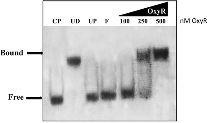
Fig 3. Electrophoretic mobility shift assay (EMSA) to indicate OxyR binding to the promoter region upstream of phnW.
Purified OxyR was added to 0.8 ng of DIG-nonradioactive labeled 199-bp DNA fragment of phnW containing the putative phnW OxyR binding domain in the binding buffer and separated on a polyacrylamide gel as described in the materials methods section. The binding reaction consisted of a labeled phnW fragment and various quantities of OxyR protein. UP is the addition of 2 μg of unrelated protein (BSA) to the binding reaction; F is free probe; the addition of increasing concentrations of OxyR (100, 250, 500 nM) to labeled phnW probe are listed; CP is the addition of 125-fold excess of unlabeled phnW DNA to the binding reaction; UD, the addition of 125-fold excess of unrelated DNA (pUCP20 plasmid) to the binding reaction. The positions of free and bound phnW probe are shown on the left (see arrows).

