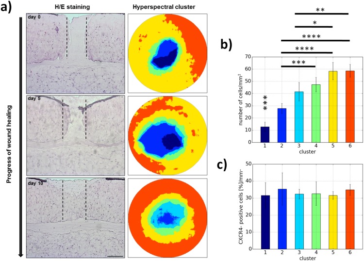
Fig 4. Histological classification of hyperspectral cluster means in relation to wound healing processes.
(a) H/E staining was performed to monitor the wound healing progress morphologically over a specific time period in vitro. The different stages were presented as false color zoomed images of the hyperspectral clusters for the wounds. (b) The quantification of the cell number in different regions of interests revealed the first time a correlation between spectral reflectance and cell quantity in the tissue. (c) Immunohistochemical investigation of CXCR4, a marker for migratory cells, determined no correlations to reflectance data. Additionally, the cells generating the characteristic hyperspectral signature did not correspond to Caspase 3-expressing apoptotic cells (not shown). Scale bar: 250 μm.

