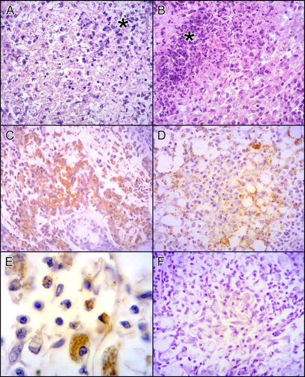
Fig 2. Lesions in BALB/c mice infected with L. (L.) amazonensis promastigotes from LV79 and PH8 strains for 12 weeks.
HE staining of BALB/c mice footpads infected with PH8 (A) and LV79 (B) with necrosis area (*). Parasite labeling in PH8 (C) and LV79 (D) lesions by IHQ using anti-Leishmania serum and anti-rabbit HRP antibody. In E, higher magnification of IHQ of PH8 lesion, in F, negative control with secondary antibody but no anti-Leishmania serum. A, B, C, D and F 400X magnification, E with 1000x magnification.

