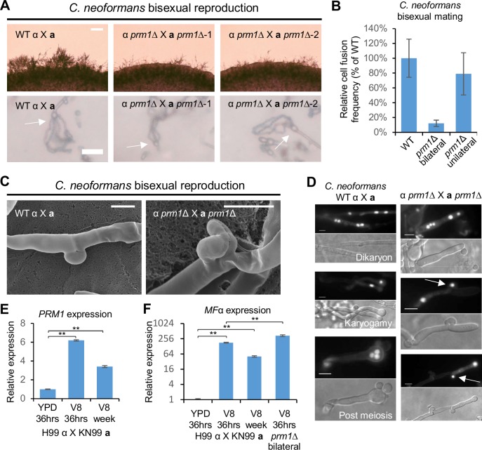
Fig 2. Deletion of PRM1 blocks cell-cell fusion and clamp cell fusion during Cryptococcus neoformans bisexual reproduction.
(A) Mating phenotypes for a wild type cross between H99α and KN99a and two independent prm1 bilateral mutant crosses between CF30 and CF448, and between CF56 and CF562. All mating patches were spotted on MS medium and incubated in the dark at room temperature. The top row shows hyphal growth on the edge of mating patches five days after inoculation. The scale bar is 100 μm. The bottom row shows the basidium and spore chain morphology (indicated by arrows)10 days after inoculation. The scale bar equals 20 μm. (B) Unilateral and bilateral prm1 mutant cell fusion frequency compared to wild type. (C) Scanning electron microscopy of clamp cell morphology of wild type cross (H99α X KN99a) and prm1 bilateral mutant cross (CF56 X CF562). The scale bar is 5 μm. (D) DAPI staining of mature hyphae and nuclei inside hyphae and basidia of C. neoformans wild type (left panel) and prm1 (right panel) bisexual crosses. Arrows in the prm1 column indicate nuclei trapped in unfused clamp cells. The scale bar is 5 μm. Gene expression patterns for (E) PRM1 and (F) MFα were examined by RT-PCR (* indicates p <0.05 and ** indicates p <0.005 for each pairwise comparison). A wild type cross (H99α X KN99a) was grown on YPD medium for 36 hours, and on V8 medium for 36 hours or one week. The prm1 bilateral mutant cross (CF56 X CF562) was grown on V8 medium for 36 hours. The Y axis for panel F is in base-2 log scale. The error bars represent the standard deviation of the mean for the three biological replicates.

