
Figure 1.
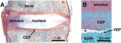
Histologic sections from human cadaveric lumbar spines. (A) Tri‐chrome Mallory–Heidenhain stained section depicting the annulus fibrosus, nucleus pulposus, vertebral bone, and cartilage endplate (CEP). (B) Safranin‐O stained section illustrating an in situ CEP avulsion (at asterisk) from the underlying vertebral endplate (VEP).