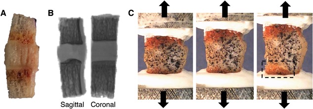
Figure 4.

Specimen is (A) cleaned of bone marrow, (B) X‐rayed in two orthogonal planes to calculate geometry, and (C) speckle‐coated on parasagittal surface, mounted in custom‐designed aluminum pots, and pulled in tension to failure. Box in (C) shows location of endplate junction failure. A, B (left), and C are all sagittal views with the inner annulus on the left and outer annulus on the right.
