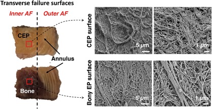
Figure 8.

(Left) Transverse failure surfaces for specimen that failed at cartilage endplate (CEP)‐bone interface at inner annulus fibrosus (AF) region and failed in mid‐annulus at outer AF region. Red squares indicate location of SEM scans; (Right) SEM scans of failure surfaces imaged for CEP; and opposing bony endplate surface.
