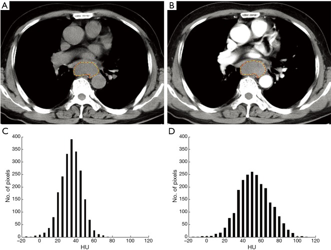
Figure 2.
A 71-year-old man with pathologically diagnosed moderately differentiated squamous carcinoma at stage IIIB (T3/N2/M0). (A) Axial unenhanced CT image shows a mass in middle of the oesophagus, and the outline indicates the ROI; (B) corresponding contrast enhanced CT image shows mild enhancement of this lesion; (C) Histogram obtained from unenhanced CT image shows pixel distribution with a bin size of 5 HU; mean =37 HU, 10th and 90th percentile =23 and 50 HU, skew =−1.57, kurtosis =51.14, and entropy =3.81, respectively; (D) Histogram obtained from contrast enhanced CT image shows; mean =55 HU, 10th and 90th percentile =35 and 77 HU, skew =1.89, kurtosis =44.65, and entropy =4.17, respectively. ROI, region of interest; HU, hounsfield units.

