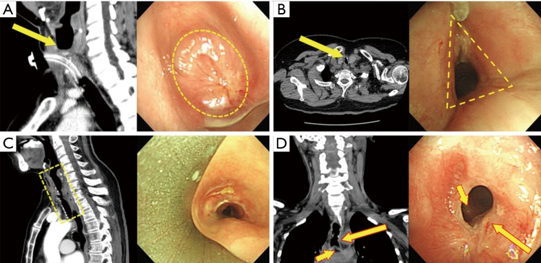
Figure 1.
CT and bronchoscopic findings according to PTTS type. (A) Subglottic type. Subglottic area was completely obstructed by fibrotic stricture (arrow and circle); (B) stoma type. Upper trachea at the tracheostomy stoma was narrowed by cartilage fracture and fibrosis (arrow). Tracheal stenosis of triangular shape was observed during the bronchoscopy (dotted triangle); (C) cuff type. Mid-trachea at the cuff level was narrowed by fibrotic bands (dotted square); (D) tip granuloma type. Distal trachea just above the carina was narrowed by fibrosis (long solid arrow: fibrotic band; short dotted arrow: carina). CT, computed tomography; PTTS, post-tracheostomy tracheal stenosis.

