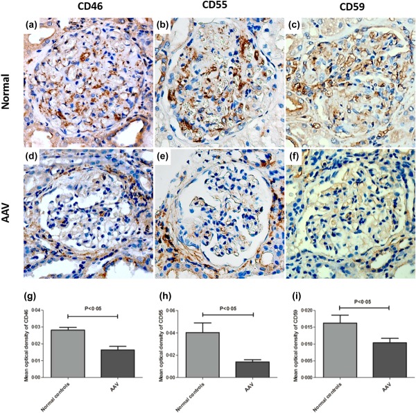
Figure 1.

Immunohistochemistry staining for CD46, CD55 and CD59 in glomeruli of renal specimens. (a–c) Immunohistochemical staining of CD46, CD55 and CD59 in glomeruli of a normal control. (d–f) Immunohistochemical staining of CD46, CD55 and CD59 in glomeruli of patient with anti‐neutrophil cytoplasmic antibody (ANCA)‐associated vasculitis. (g–i) Comparison of the mean optical density of CD46, CD55 and CD59 in glomeruli of patients with ANCA‐associated vasculitis to that of normal controls. [Colour figure can be viewed at wileyonlinelibrary.com]
