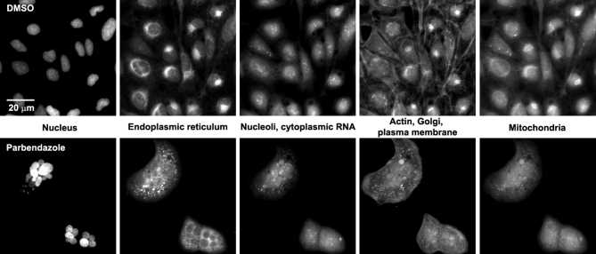
Figure 1:
Sample images of U2OS cells from the small-molecule Cell Painting experiment. Images are shown from a DMSO well (negative control, top row) and a parbendazole well (bottom row). The columns display the 5 channels imaged in the Cell Painting assay protocol (see Table 1 for details about the stains and channels imaged).

