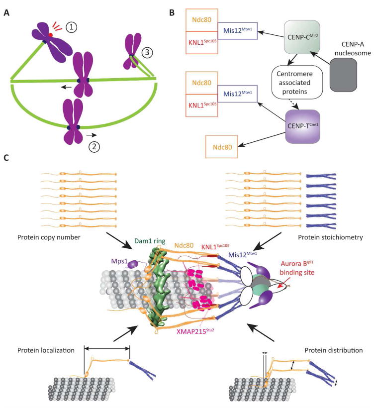
Figure 1. The function and protein architecture of the kinetochore.
A Cartoon of a mitotic spindle displaying the three main kinetochore functions: (1) Activation of the Spindle Assembly Checkpoint, (2) generation of bidirectional chromosome movement that is coupled to microtubule polymerization and depolymerization, and (3) correction of monopolar attachment of sister kinetochores. B (left to right = microtubule plus-end to centromere) The conserved, dual pathways (solid arrows – direct interaction, dashed arrow – indirect interaction) that assemble the KMN network, which forms the interface of the kinetochore with the microtubule plus-end. C Reconstruction of the protein architecture of the budding yeast kinetochore using fluorescence microscopy measurements and protein structures [7, 8, 14, 15, 32, 33, 48–50, 73]. Centromere-associated proteins are represented by white, oblong shapes.

