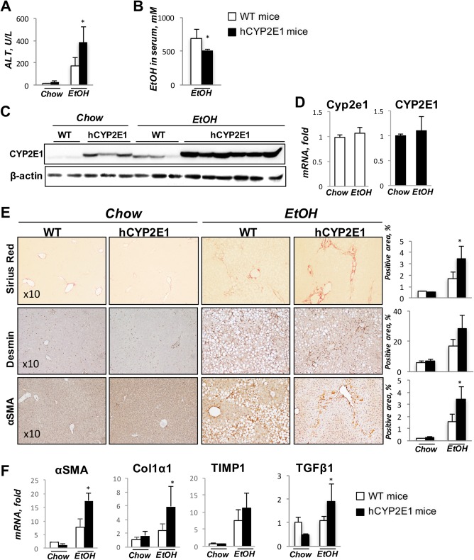
Figure 1.

Hepatic fibrosis is increased in hCYP2E1 mice after iG alcohol feeding. Wt and hCYP2E1 mice were subjected to iG alcohol feeding with weekly binges for 8 weeks. Wt and hCYP2E1 littermates fed with normal chow were used as controls. (A) Liver function was assessed by ALT. (B) Ethanol concentration in serum was measured in Wt and hCYP2E1 mice after iG alcohol feeding. (C) Immunoblotting of CYP2E1 in liver. (D) CYP2E1 mRNA expression in both Wt and hCYP2E1 liver. (E) Representative images of Sirius Red staining, immunohistochemistry staining of Desmin, and α‐SMA. The stained area was shown with quantification of morphometric analysis of each staining. (F) Hepatic expression of fibrogenic genes was measured by mRNA‐fold induction level in Wt and hCYP2E1 livers. The P value was measured between Wt and hCYP2E1 with iG alcohol feeding; the data were shown as mean ± SEM; *P < 0.05. Abbreviations: EtOH, ethanol; iG, intragastric.
