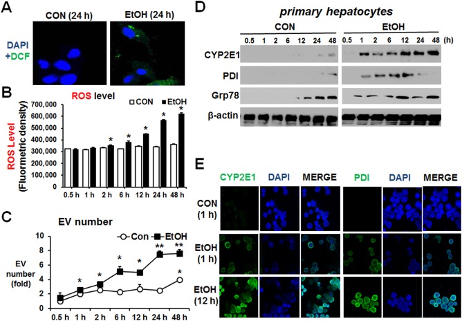
Figure 6.

Alcohol elevated EV number through increased oxidative and ER stress in primary hepatocytes. Confluent mouse primary hepatocytes were treated with 100 mM ethanol for 0.5, 1, 2, 6, 12, 24, and 48 hours. (A) Confocal images of increased ROS determined by green dichlorofluorescein‐diacetate fluorescence. (B) ROS levels are shown after fluorometric measurement of dichlorofluorescein‐formaldehyde in the hepatocyte lysates collected at the indicated times after exposure to 100 mM ethanol. (C) Total number of EVs in each group was measured by Nanosight. *P < 0.05, **P < 0.01. (D) Immunoblot analyses for CYP2E1, PDI, and Grp78 in the indicated hepatocyte lysates. β‐Actin, used as a loading control, is shown. (E) Confocal images of CYP2E1 and PDI were increased at 12 hours after alcohol exposure. Abbreviations: CON, control; DAPI, 4′,6‐diamidino‐2‐phenylindole; EtOH, ethanol. Data are shown as means ± SD.
