
Figure 3.
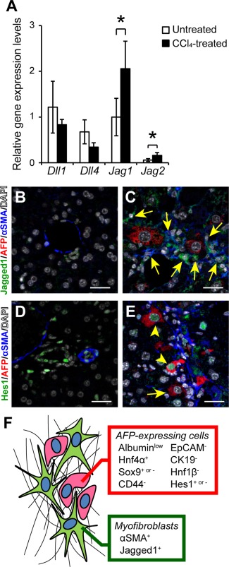
Activation of Notch signaling in AFP‐positive cells by the adjacent Jagged1‐expressing myofibroblasts. (A) Relative gene expression levels of Delta‐like 1 (Dll1), Delta‐like 4 (Dll4), Jagged1 (Jag1), and Jagged2 (Jag2) were compared between untreated and CCl4‐treated fibrotic liver tissues. The values are the means ± SD from four mice (two males, two females) in each group. An asterisk indicates that the difference between groups was statistically significant (P < 0.05). Expression of Jagged1, AFP, and αSMA was examined by immunofluorescent staining in (B) untreated and (C) CCl4‐treated liver tissues. The arrows indicate myofibroblasts co‐expressing Jagged1 and αSMA. Scale bars, 50 μm. Expression of Hes1, AFP, and αSMA was also examined in (D) untreated and (E) CCl4‐treated liver tissues. The arrowheads and arrow indicate AFP‐positive cells with and without co‐expression of Hes1, respectively. Scale bars, 50 μm. (F) Schematic representation of AFP‐positive cells located adjacent to Jagged1‐expressing activated myofibroblasts in fibrotic liver. Abbreviation: DAPI, 4´,6‐diamidino‐2‐phenylindole.