
Figure 5.
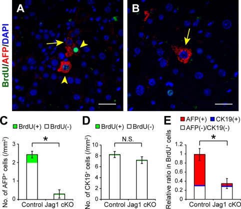
Suppression of mobilization and proliferation of AFP‐positive cells after partial hepatectomy in CCl4‐treated Jagged1 cKO mice. Cell proliferation was evaluated by BrdU staining in regenerating fibrotic liver 48 hours after partial hepatectomy. BrdU (100 μmol/kg body weight) was injected intraperitoneally 2 hours before excising the liver. Costaining of AFP and BrdU together with DAPI nuclear staining was performed using liver specimens obtained from (A) CCl4‐treated control and (B) Jag1 cKO mice. The arrowheads and arrows indicate AFP‐expressing cells with and without BrdU costaining, respectively. Scale bars, 100 μm. The total numbers of (C) AFP‐positive or (D) CK19‐positive cells with or without BrdU co‐expression and (E) the relative ratio of each cell population in BrdU‐positive cells were compared between control and Jag1 cKO mice as described in Fig. 1. The values indicate the means ± SD from three mice in each group (two males, one female). An asterisk indicates that the difference between groups was statistically significant (P < 0.01). Abbreviations: DAPI, 4´,6‐diamidino‐2‐phenylindole; N.S., not significant.