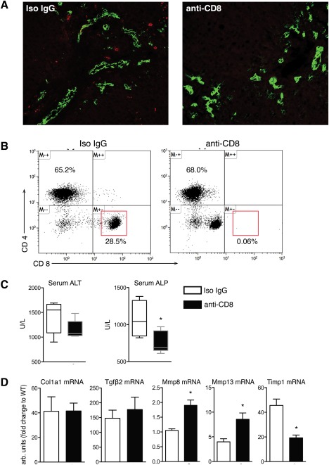
Figure 3.

CD8+ T‐cell depletion ameliorates liver injury and has beneficial effects on fibrosis‐related gene expression. (A) Immunofluorescent staining of CD8+ (red) and bile ducts visualized by pan‐CK (green) in liver sections from male Mdr2–/–;CD39–/– mice 3 days after administration of anti‐CD8 monoclonal antibody or the respective isotype control (original magnification, ×200). (B) Flow cytometry analysis of CD8 expression in splenocytes isolated from Mdr2–/–;CD39–/– mice treated for 3 days with anti‐CD8 or isotype control. Cells were gated on CD3+ subsets. (C) CD8+ T‐cell depletion resulted in a significant decrease in serum ALT and ALP. (D) CD8+ T‐cell depletion led to a profibrolytic shift in gene expression with significantly reduced TIMP‐1 expression and increased MMP‐8 and MMP‐13 expression. Data are mean ± SEM (n = 3‐5 mice/bar). *P < 0.05 compared to isotype‐treated control mice (t test). Abbreviations: CK, cytokeratine; IgG, immunoglobulin G; Tgfβ2, transforming growth factor β2; TIMP‐1, tissue inhibitor of metalloproteinase 1.
