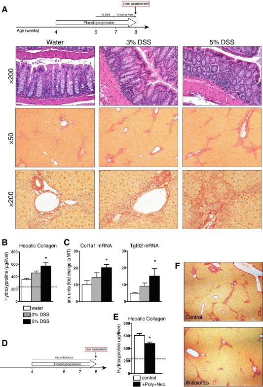
Figure 4.

Intestinal bacteria promote sclerosing cholangitis in Mdr2–/– mice. (A) Experimental design of experimental colitis model in 6‐week‐old male FVB.Mdr2–/– mice. After 7 days of DSS administration, colitis was assessed, the DSS‐supplemented water was replaced by normal drinking water, and livers assessed 7 days later. Hematoxylin and eosin staining of colons (×200) and Sirius Red staining of liver specimens from Mdr2–/– mice receiving drinking water (left), 3% DSS (middle), or 5% DSS (right) for 7 days. Magnification ×50 (middle panel) and ×200 (lower panel, portal area). (B) Hydroxyproline content in livers from control and DSS‐treated Mdr2–/– mice. (C) Hepatic expression of Col1a1 and Tgfβ2. (D) Schematic outline for the antibiotic treatment in male Mdr2–/– mice. (E) Hepatic hydroxyproline content and (F) representative connective tissue stain (Sirius Red, ×50) in livers from 8‐week‐old Mdr2–/– mice after 4 weeks of antibiotic treatment with polymyxin B (100 mg/kg) + neomycin (220 mg/kg). Data are shown as mean ± SEM (n = 4‐5 mice/bar). *P < 0.05 compared to age‐matched Mdr2–/– controls (analysis of variance followed by Dunnett's posttest). Abbreviations: DSS, dextran sulfate sodium; FVB, Friend virus B‐type; Tgfβ2, transforming growth factor β2.
