
Figure 4. Image segmentation in CDI iCell neurons.
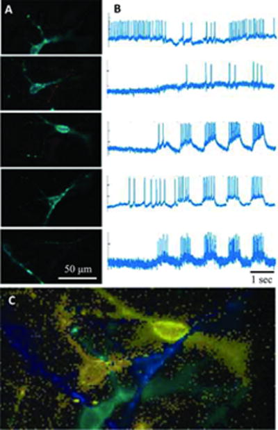
At high magnification, neurons can be identified manually, but at lower magnification or in high-density cultures, automated analysis is required. Principal component analysis (PCA) identifies significant sources of variation in the movie, representative of neuronal firing, but time traces and pixel weight maps are typically mixtures of multiple neurons. Independent component analysis (ICA) “unmixes” the signal, identifying individual time traces even in crowded images where cells are partially overlapped. This figure shows results from one such field of view, where 5 overlapped neurons are present. (A) Identified neuronal sources and (B) their action potential trains resulting from PCA/ICA analysis. Red indicates negative pixel weights. (C) The composite image with all sources combined.