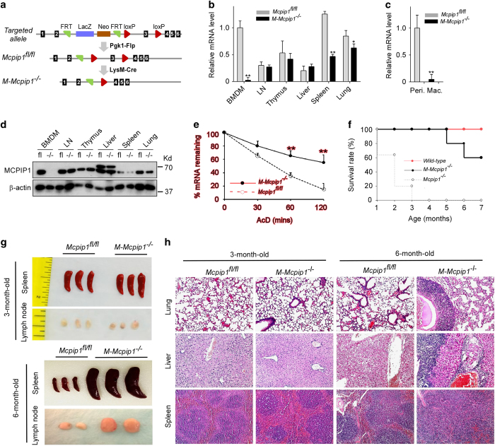
Figure 1.
Generation and characterization of M-Mcpip1−/− mice. (a) Schematic map of the strategy used for generating the Mcpip1 conditional targeted allele, Mcpip1floxflox, and M-Mcpip1−/− mice. (b) qPCR analysis of the MCPIP1 mRNA levels in BMDMs and the indicated tissues collected from Mcpip1flfl and M-Mcpip1−/− mice. Data are representative of three independent experiments with 3 mice per group. *P<0.05, **P<0.001 by Student’s t-test. (c) qPCR analysis of the MCPIP1 mRNA levels in peritoneal macrophages collected from Mcpip1flfl and M-Mcpip1−/− mice. Data are representative of three independent experiments with 3 mice per group. **P<0.001 by Student’s t-test. (d) Immunoblot analysis of the MCPIP1 protein levels in BMDMs and indicated tissues from Mcpip1flfl and M-Mcpip1−/− mice. β-actin served as a loading control. Data are representative of three independent experiments. (e) BMDMs from Mcpip1flfl and M-Mcpip1−/− mice were stimulated with LPS (1 μg/ml) for 4 h and then incubated with AcD (5 μg/ml) for different time points as indicated. The mRNA levels of IL-6 were measured by qPCR and normalized as 100% at the 0-hr time point. Data were presented as the means±s.d., n=4. (f) Survival rate of Mcpip1 global knockout (Mcpip1−/−) mice, myeloid-specific Mcpip1 knockout (M-Mcpip1−/−) mice and wild-type mice (n=10). (g) Photographs of spleens and lymph nodes collected from Mcpip1flfl and M-Mcpip1−/− mice at 3 months and 6 months of age. (h) Hematoxylin-and-eosin staining of lung and liver sections from Mcpip1flfl and M-Mcpip1−/− mice at 4 months of age or 6 months of age, respectively. Original magnification is 100×. Data are representative of three independent experiments.

