Fig. 1.

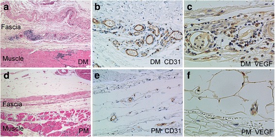
The histological images of fasciitis, CD31-expressing blood vessels and vascular endothelial growth factor (VEGF)-expressing cells in the fascia of patients with dermatomyositis (DM) or polymyositis (PM). a Fasciitis in a patient with DM. H&E staining shows massive inflammatory cell infiltration around the small blood vessels and hyperplasia of the fascia (original magnification × 50). b Immunohistochemical staining shows the proliferation of CD31-expressing capillaries and venules (brown) in the fascia of a patient with DM (original magnification × 200). c Immunohistochemical staining shows the accumulation of VEGF-expressing cells (brown) around the blood vessels in the fascia of a patient with DM (original magnification × 400). d No fasciitis is observed in a patient with PM (original magnification × 50). e Immunohistochemical staining for CD31 (brown) shows no angiogenesis in the fascia of a patient with PM (original magnification × 200). f Immunohistochemical staining shows a small number of VEGF-expressing cells (brown) in the fascia of a patient with PM (original magnification × 400)
