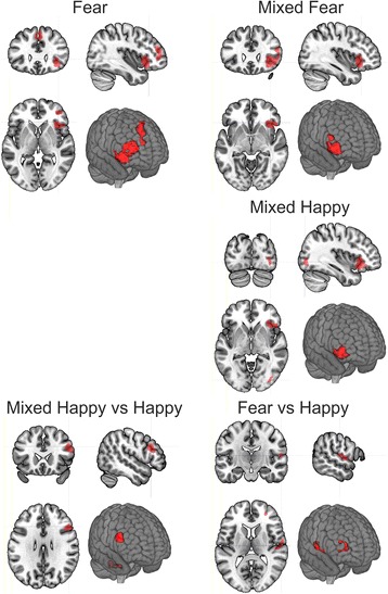
Fig. 2.

Between group differences in brain activation to fear, mixed fear, and mixed happy stimuli respectively.1 Fear: Right Middle Frontal Gyrus (RMFG), Right Anterior Insula (RAI), Right Ventrolateral Prefrontal Cortex (VLPFC), Right and Left Medial Frontal Gyrus, Dorsomedial Prefrontal Cortex (DMPFC). 2 Mixed Fear: Right Anterior Insula (RAI), Right Ventrolateral Prefrontal Cortex (VLPFC). 3 (Happy: no between group difference). 4 Mixed Happy: Right Anterior Insula (RAI), Right Ventrolateral Prefrontal Cortex (VLPFC), Right Middle Occipital Gyrus (RMOG)
The clusters corresponding to the interactions between stimulus condition and study group are also presented. 1 Fear vs. Happy: Right Superior Temporal Gyrus (RSTG), Right Rolandic Operculum (RRO), Right Middle Frontal Gyrus (RMFG). 2 Mixed Happy vs. Happy:, Vermis of the Cerebellum Right Middle and Inferior Frontal Gyrus, Right Dorsolateral Prefrontal Cortex (DLPFC). Results are based on permutation based statistics (n = 10,000) from Statistical non-Parametric Mapping (SnPM).
