Fig. 2.

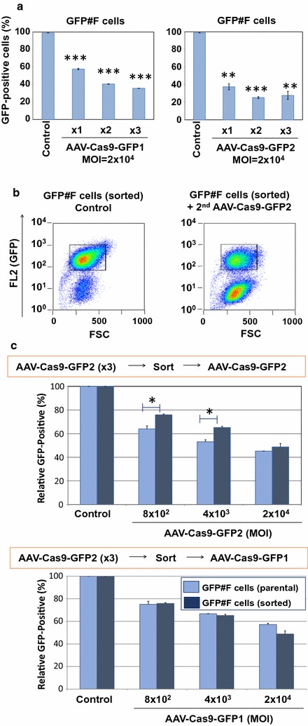
a GFP#F cells were transduced with two AAV vectors for three consecutive days at MOI 2 × 10⁴. When treated with AAV-Cas9-GFP1 (left panel), 57.7 ± 0.7, 40.6 ± 0.2 and 35.6 ± 0.1% cells were found GFP-positive, while 37.5 ± 3.3, 25.2 ± 1.1, 27.7 ± 4.5% of cells were found GFP-positive with AAV-Cas9-GFP2 (right panel) vector infection after one, two or three administrations, respectively. Error bars indicate STDEV. *P < 0.05, **P < 0.01, and ***P < 0.001. b We sorted the GFP-positive cell population in the GFP#F cells, which were treated by AAV-Cas9-GFP2 vector at MOI 2 × 10⁴ for three consecutive days. Sorted cells were used for AAV-Cas9-GFP vector superinfection study. Representative FACS images for sorted cells (left panel) and AAV-superinfected cells (right panel) were shown. c Parental GFP#F cells and sorted cells were transduced with AAV-Cas9-GFP2 at MOI of 8 × 10², 4 × 10³, 2 × 10⁴ gc and GFP-positive cell populations were analyzed by FACS (upper panel). Lower panel shows the results of GFP-positive cell populations after super-infection with another GFP-targeting vector, AAV-Cas9-GFP1 vector. The averages of two independent experiments are shown. Error bars represent SEM (*P < 0.05)
