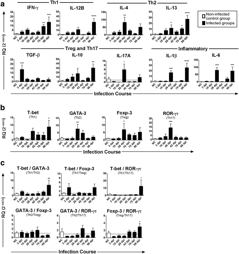
Fig. 2.

Temporal changes of the mRNA expression of cytokines and transcription factors in the liver of buffaloes experimentally infected with Fasciola gigantica. The X axis represents days post infection (dpi), control animals are represented by empty bars and Y axis represents the mRNA relative expression of target genes relative to GAPDH based on 2–ΔΔCq calculation. Columns show the means and error bars show SEMs. The log number of mRNA relative expression of a IFN-γ, IL-1β, IL-4, IL-6, IL-10, IL-12B, IL-13, IL-17A, TGF-β and b T-bet, GATA-3, Foxp3, and ROR-γτ, are shown. c Trends in the ratios between each pair of transcription factors during the course of infection, which was used as indicators of the balance between Th1/Th2 and Treg/Th17. Significant differences of each time-point compared with control non-infected (NC) group: *P < 0.05; **P < 0.01; ***P < 0.001 or ****P < 0.0001 (analyzed by one-way ANOVA, post-hoc LSD test)
