
Figure 1.
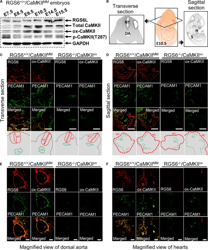
Expression of Regulator of G protein signaling 6 (RGS6) and oxidized Ca2+/calmodulin‐dependent protein kinase II (ox‐CaMKII) in mouse embryos. A, Western blot analysis of RGS6, ox‐CaMKII, phosphorylated CaMKII (p‐CaMKII), and total Ca2+/calmodulin‐dependent protein kinase II (CaMKII) expression in RGS6+/+/CaMKIIMM embryos at different gestation periods. B, Schematic illustration of transverse and sagittal sections of embryos used for immunostaining. C, Immunohistochemical analyses of RGS6 and ox‐CaMKII (red) expression in the dorsal aorta of embryos (transverse section) at E10.5 and co‐localization with the endothelial marker platelet endothelial cell adhesion molecule 1 (PECAM1) (green, upper). The schematic view showing the location of the neural tube (NT) and dorsal aorta (DA), in the respective pictures, are indicated by dotted red lines and green lines, respectively (lower). D, Immunohistochemical analysis of RGS6 and ox‐CaMKII (red) expression in the hearts of embryos (sagittal section) at E10.5 and co‐localization with PECAM1 (green, upper). The schematic view showing the location of the myocardium (MC) and endocardium (EC), for the respective pictures, are indicated by dotted red lines and green lines, respectively (lower). E, Co‐localization of RGS6 and ox‐CaMKII (red) with PECAM1 (green) in the DA region at higher magnification from embryo sections at E10.5. F, Co‐localization of RGS6 and ox‐CaMKII (red) with PECAM1 (green) immunostaining of endothelial cells in heart at higher magnification from E10.5 embryo sections. Decreased numbers of positive PECAM1‐stained endothelial cells were found in RGS6−/−/CaMKIIVV embryos compared with RGS6+/+/CaMKIIMM embryos. Scale bars, 50 μm (C and D) and 20 μm (E and F). H indicates heart; S, somites.