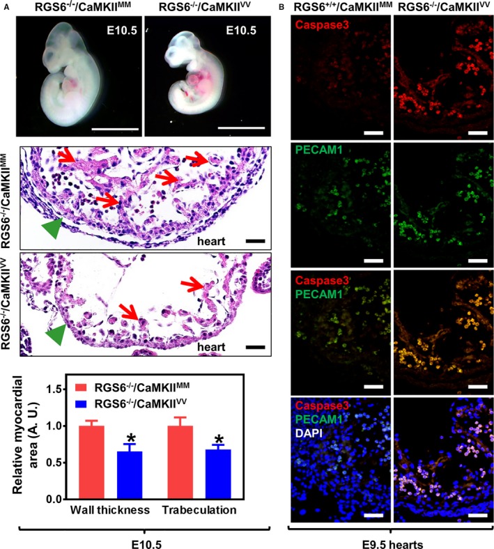
Figure 2.

Cardiovascular abnormalities in Regulator of G protein signaling 6 (RGS6)−/−/Ca2+/calmodulin‐dependent protein kinase II (CaMKII)VV embryos. A, Representative images of freshly dissected live E10.5 embryos (upper panel, scale bar=200 μm) and hematoxylin and eosin (H&E) staining of heart sections taken from E10.5 embryos (middle panel, scale bar=20 μm). Green arrowheads indicate normal (RGS6−/−/CaMKIIMM) and thinned (RGS6−/−/CaMKIIVV) myocardium and red arrows indicate regions of normal trabeculation in RGS6−/−/CaMKIIMM hearts and its loss in RGS6−/−/CaMKIIVV hearts of H&E‐stained sections (middle panel). Both myocardial thickness and the level of trabeculation were significantly reduced in RGS6−/−/CaMKIIVV embryos when compared with RGS6−/−/CaMKIIMM embryos, analyzed by Mann–Whitney test (lower panel). N=7 to 9 embryos, *P<0.05. B, Increased caspase 3 (red) and platelet endothelial cell adhesion molecule 1 (PECAM1) (green)–stained double‐positive cells (yellow) in the heart ventricle of RGS6−/−/CaMKIIVV embryos at E9.5 compared with RGS6−/−/CaMKIIMM. Scale bar=20 μm. DAPI indicates 4′,6‐diamidino‐2‐phenylindole.
