
Figure 5.
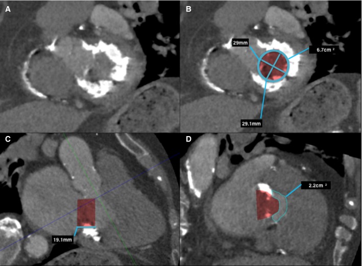
Predicting neo–left ventricular outflow tract (LVOT) area in a native mitral valve with severe mitral annular calcification on postcontrast cardiac computerized tomography (CT) using 30 mL iodinated contrast. A, Left ventricle short‐axis multiplanar reformat (MPR) at the level of the mitral annulus with severe mitral annular calcification, with (B) mitral annular measurements and segmented valve area. Three‐chamber MPR (C) demonstrates the proposed valve prosthesis, in this case a 29‐mm Edwards SAPIEN XT valve. Planimetry of the neo‐LVOT (C, blue line), with planimetry of the neo‐LVOT performed in an orthogonal plane (D).