Figure 2.

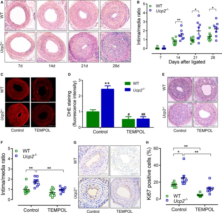
Uncoupling protein 2 (UCP2) ablation exacerbates mouse carotid artery ligation‐induced myointimal hyperplasia. A, Hematoxylin and eosin (H&E)‐stained sections of ligated carotid arteries from Ucp2 −/− mice and wild‐type (WT) littermates 7, 14, 21, and 28 days after ligation. Bar=50 μm. B, Intima/media ratio of ligated arteries. n=8. *P<0.05, **P<0.01. C, Dihydroethidium (DHE)‐stained frozen sections of ligated carotid arteries from Ucp2 −/− and WT mice treated with or without 4‐hydroxy‐2,2,6,6‐tetramethyl‐piperidinoxyl (TEMPOL) in drinking water (1 mmol/L) for 21 days. Bar=50 μm. D, Quantification of DHE fluorescence intensity. n=6. **P<0.01 vs WT‐control; # P<0.05, ## P<0.01 vs isogenic mice treated without TEMPOL. E, H&E‐stained sections of ligated carotid arteries from mice treated with or without TEMPOL. Bar=50 μm. F, Intima/media ratio of ligated arteries. n=8 in each group. **P<0.01. G, Immunohistochemistry staining of Ki67 (brown) in sections of ligated carotid arteries from mice treated with or without TEMPOL. Bar=50 μm. H, Percentage of Ki67‐positive cells within neointima. n=8. *P<0.05, **P<0.01.
