Figure 1.

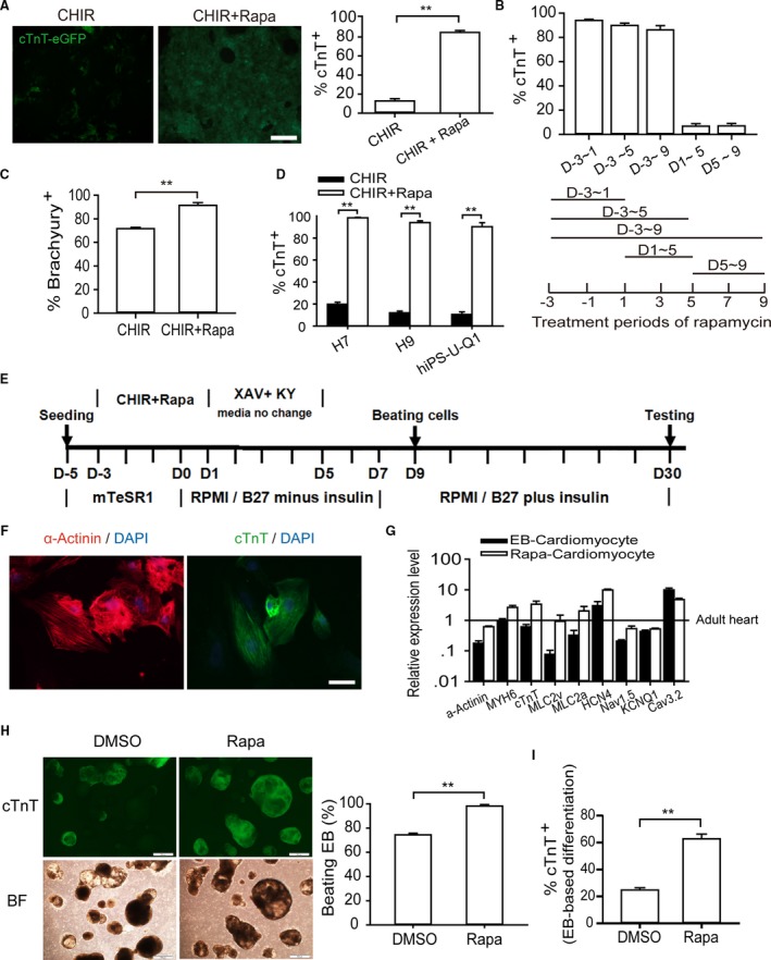
Rapamycin promotes cardiomyocyte differentiation in monolayer‐based growth factor–free system. A, Analyzing the efficiency of cardiomyocyte differentiation in terms of reporter activity. Left and right graphs show the ratio of GFP‐positive cardiomyocytes identified by fluorescence observation and flow cytometry analysis (12 μmol/L CHIR, 10 nmol/L rapamycin). The asterisks indicate statistically significant differences compared with controls (n=5; **P<0.01). Scale bars, 200 μm. B, Effective time window of rapamycin treatment. Addition of rapamycin from day −3 to day 1 is sufficient for highly efficient cardiogenesis based on GFP‐positive cell percentage (n=5). C, Flow cytometry validation of Brachyury‐positive cells at day 4 (n=5; **P<0.01). D, hESC lines H9 and H7 and hiPSC line U‐Q1 were induced into cardiomyocytes with or without rapamycin (n=6; **P<0.01). E, Schematic of protocol for small molecule–mediated cardiomyocyte differentiation from hPSC. F, Immunostaining of cTnT and α‐actinin showed sarcomere organization. Scale bars, 10 μm. G, Gene expression analysis of rapamycin‐induced cardiomyocytes. The expression levels of cardiac marker genes (α‐Actinin,MYH6, cTnT,MLC2v, and MLC2a) and channel genes (HCN4, Nav1.5,KCNQ1, and Cav3.2) were nearly equal to those of EB‐derived cardiomyocyte and adult heart tissue, which was considered to be 100% (n=5). H and I, The effect of rapamycin on EB‐based cardiac differentiation. Rapamycin was added in the first stage (0 to 4 days) of EB differentiation, and cardiac differentiation efficiency was assessed at day 15. The percentage of beating EB (H) and the ratio of GFP‐positive cardiomyocytes (I) were analyzed (n=5; **P<0.01). Scale bars, 500 μm. BF indicates bright field; CHIR, CHIR99021; DMSO, dimethyl sulfoxide; EB, embryoid body; eGFP, enhanced green fluorescent protein; hESC, human embryonic stem cell; KY, KY02111; Rapa, rapamycin; XAV, XAV939.
