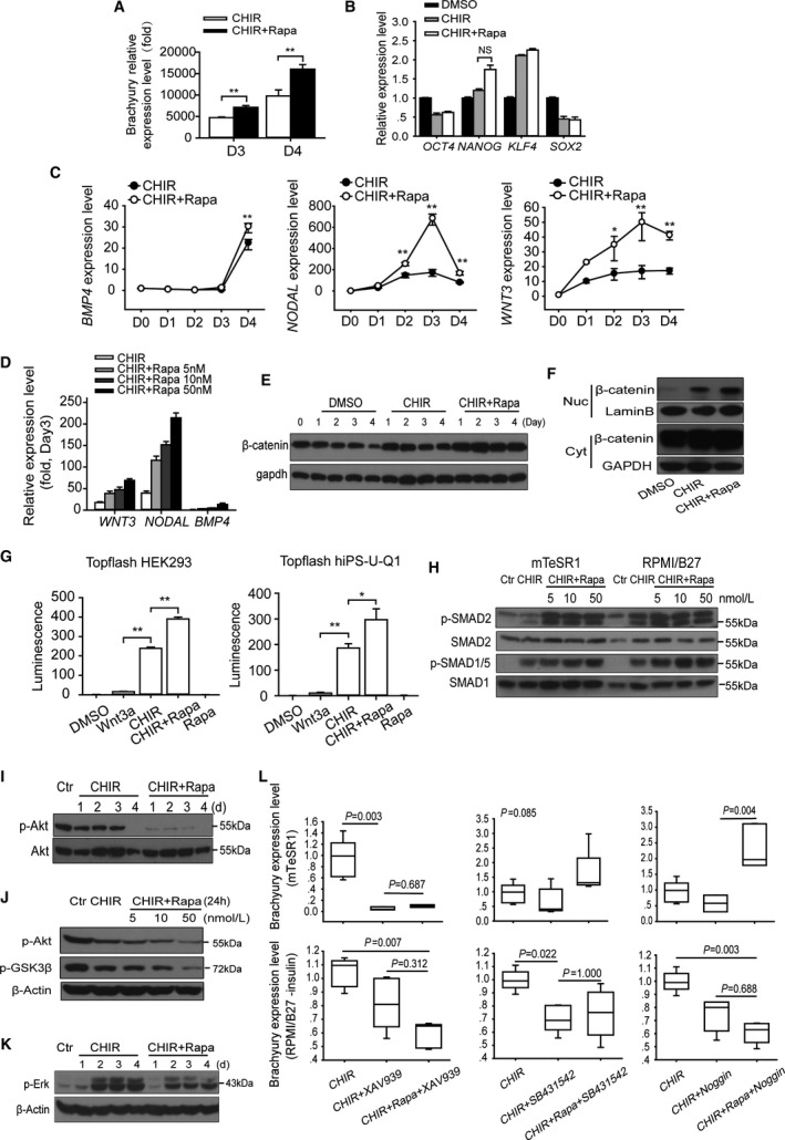
Figure 6.

Rapamycin promotes multiple signaling pathways essential for mesoderm induction. A, The transcriptional level of Brachyury with or without rapamycin treatment in mTeSR1 (D3) and RPMI B27 (D4) medium (n=5; **P<0.01). B, The transcriptional level of hESC self‐renewal core factors, including OCT4,NANOG,KLF4, and SOX2 with or without rapamycin treatment (n=5; NS means no significant difference). C, Daily transcriptional levels of endogenous growth factors including BMP4,NODAL, and WNT3 in H9 hESCs induced by CHIR with or without rapamycin (10 nmol/L). Treated cells were collected and analyzed at indicated time points, and 1‐way ANOVA followed by post hoc Bonferroni test was used to calculate significant difference between groups at each time point (n=4; *P<0.05, **P<0.01). D, Quantitative real‐time PCR analysis of WNT3,NODAL, and BMP4 expression with different concentrations of rapamycin at day 3 (n=5). E, The expression of β‐catenin with or without rapamycin treatment. F, Nucleocytoplasmic separation experiment showed the distribution of β‐catenin, which reflects the activity of Wnt signaling. G, Topflash reporter assay performed using hiPS‐U‐Q1 and HEK293T cells. The effect of CHIR, rapamycin, or DMSO was examined, and Wnt3a (100 ng/mL) was added as a positive control (n=5; *P<0.05, **P<0.01). H, Expression of phosphorylation of SMAD2 (Ser467), SMAD1/5 (Ser463/465), and total SMAD2 and SMAD1/5 were analyzed via Western blots in H9 cells treated with 12 μmol/L CHIR or CHIR plus 10 nmol/L rapamycin for 6 hours. I, The activity of PI3K/AKT signaling with or without rapamycin treatment was tested by the phosphorylation of AKT at Ser473 via Western blot with total AKT expression as reference. J, Western blot analysis shows that the suppression effect of rapamycin on the Akt pathway was concentration dependent, and the level of GSK‐3β (Ser9) phosphorylation, which is an index of Akt activity, had a similar change. K, The activity of FGF‐Erk signaling pathway was shown by the Erk (Ser44/42) phosphorylation with or without rapamycin treatment at different time points. L, Analyzing the influences of inhibition of Wnt, NODAL, and BMP4 signaling pathways on rapamycin‐induced Brachyury expression in mTeSR1 and RPMI/B27 conditions (n=5; Kruskal‐Wallis test followed by Dunn post hoc comparison). CHIR indicates CHIR99021; Ctr, control; Cyt, cytoplasm; DMSO, dimethyl sulfoxide; hESC, human embryonic stem cell; Nuc, nucleus; PCR, polymerase chain reaction; Rapa, rapamycin.
