
Figure 2.
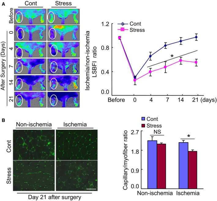
Chronic stress impaired blood flow recovery and capillary formation in ischemic muscles of the mice. A, Representative laser speckle perfusion imaging showed a low perfusion signal (dark blue) in the ischemic hindlimbs of stressed mice and a high signal (red) in control mice. The ratio of ischemic to nonischemic laser speckle blood flow was lower in the stressed mice than in the nonstressed control mice during the follow‐up period. Data are mean±SE (n=6). *P<0.01 by 2‐way repeated‐measures ANOVA and Bonferroni post hoc tests. B, Fluorescent staining was performed using tomato lectin to visualize capillaries in ischemic and nonischemic thigh adductor muscle. Quantitative data showed the reduction of capillary density as expressed by the capillary‐to‐myofiber ratio in ischemic muscle of the stressed mice (n=6). Data are mean±SE. *P<0.05, NS, not significant by Student unpaired t test or ANOVA and Tukey's post hoc tests. Scale bar, 50 μm. LSBFI indicates laser speckle blood flow imaging.