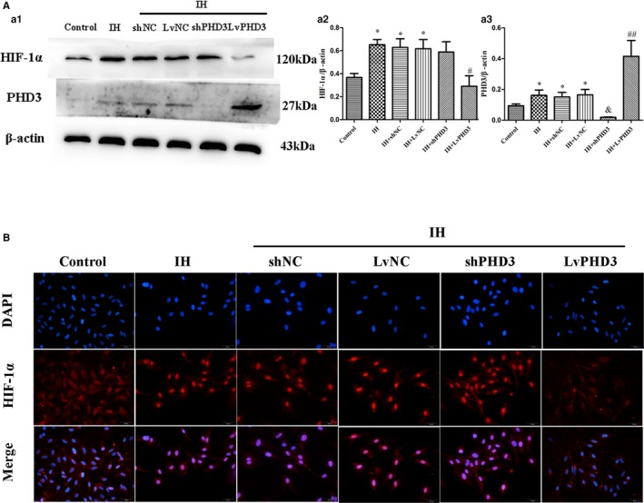
Figure 7.

PHD3 overexpression mitigates IH‐induced HIF‐1α activation in endothelial cells. A, Western blot analysis and quantification of HIF‐1α (a1 and a2), PHD3 (a1 and a3). B, Representative images of immunofluorescence staining with antibodies to HIF‐1α (red). Nuclei were counterstained with DAPI (blue; original magnification, ×400; bars=20 μm). *P<0.05 vs control; # P<0.05 vs IH+LvNC; ## P<0.01 vs IH+LvNC; & P<0.05 vs IH+shNC. DAPI indicates 4',6‐diamidino‐2‐phenylindole; HIF‐1, hypoxia‐inducible factor 1 alpha; IH, intermittent hypoxia; LvPHD3, lentivirus carrying PHD3 cDNA; PHD3, prolyl 4‐hydroxylase domain protein 3; shPHD3, short‐hairpin PHD3.
