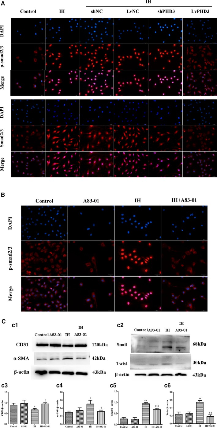
Figure 8.

PHD3 overexpression mitigates IH‐induced smad2/3 activation in endothelial cells. A, Representative images of immunofluorescence staining with antibodies to p‐smad2/3 (red) and smad2/3 (red). Nuclei were counterstained with DAPI (blue; original magnification, ×400; bars=20 μm). B, Representative images of immunofluorescence analysis of p‐smad2/3 (red) localization in HUVECs with or without IH or A83‐01 treatment. Nuclei were counterstained with DAPI (blue; original magnification, ×400; bars=20 μm). C, Western blot analysis and quantitative of CD31 (c1 and c3), α‐SMA (c1 and c4), Snail (c2 and c5), and Twist (c2 and c6). *P<0.05 vs control; **P<0.01 vs control; # P<0.05 vs IH+LvNC; ## P<0.01 vs IH+LvNC. DAPI indicates 4',6‐diamidino‐2‐phenylindole; HIF‐1, hypoxia‐inducible factor 1 alpha; HUVECs, human umbilical vein endothelial cells; IH, intermittent hypoxia; LvPHD3, lentivirus carrying PHD3 cDNA; PHD3, prolyl 4‐hydroxylase domain protein 3; p‐ smad2/3, phosphorylated small mothers against decapentaplegic 2 and 3; shPHD3, short‐hairpin PHD3; α‐SMA, alpha‐smooth muscle actin.
