Figure 5.
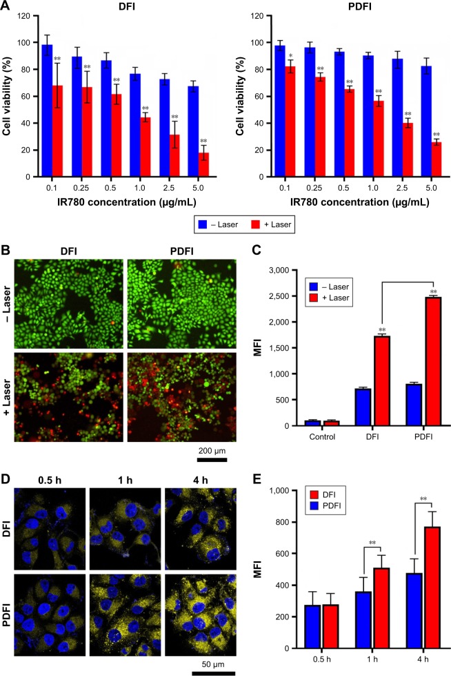
The cytotoxicity and uptake of PDFI nanoparticles in MHCC-97H cells.
Notes: (A) The cytotoxicities of DFI nanocores and PDFI nanoparticles after irradiation with (+) and without (−) an 808-nm laser at a powder density of 2 W/cm2 for 2 min. *p<0.05 and **p<0.01 for comparing treatments with and without laser irradiation. (B) The fluorescence microscopy images of cells with various treatments. The concentration of IR780 was 2.5 μg/mL. The live cells showing green fluorescence were stained with calcein-AM and the dead cells showing red fluorescence were stained with ethidium homodimer-1. (C) The comparison of MFIs of intracellular DCF produced from the reaction of DCFH-DA with ROS after various treatments. (D) The confocal images of cells incubated with DFI nanocores and PDFI nanoparticles for different times at the IR780 concentration of 2.5 μg/mL. (E) The comparison of MFIs of IR780 in cells after various treatments. **p<0.01 for comparing two treatment groups.
Abbreviations: DCF, 2′,7′-dichlorofluorescein; DCFH-DA, 2′-7′-dichlorodihydrofluorescein diacetate; DFI, IR780-loaded DOPE/PF68 complex; DOPE, 1,2-dioleoyl-sn-glycero-3-phosphoethanolamine; MFI, mean fluorescence intensity; PDFI, pullulan coated DFI nanocore; PF68, Pluronic F68; ROS, reactive oxygen species.

