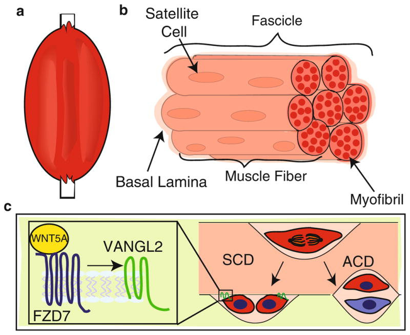
Fig. 16.2.

WNT/PCP signals during SCD in satellite stem cells. (a) Cartoon of a limb comprising skeletal muscle and bone. (b) Skeletal muscle composed of seven fascicles, each of which contains muscle fibers (seven depicted here). Within each muscle fiber are multiple myofibrils. The satellite stem cells (red) reside in between the encapsulating basal lamina and the sarcolemma (not shown) of each muscle fiber. (c) Satellite stem cells undergo both SCD and ACD. The left arrow indicates the outcome of an SCD where both daughter cells are in contact with the sarcolemma anchored by VANGL2 (green). The right arrow indicates an ACD in which only one daughter cell is in contact with the sarcolemma, remaining a satellite stem cell (red), and the other daughter contacts the basal lamina, becoming a progenitor cell (blue). The enlarged inset depicts WNT5A (yellow) bound to FZD7 (blue) during an SCD, triggering the accumulation of VANGL2 on opposite poles of the daughter cells
