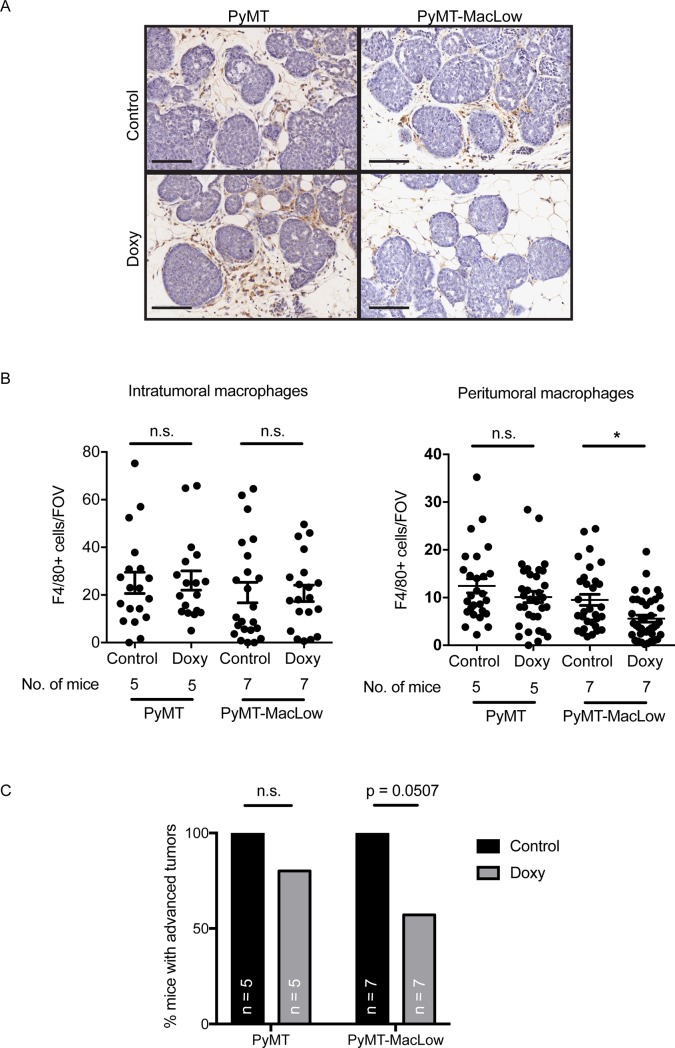
Fig 2. Doxycycline reduces the number of macrophages surrounding mammary tumors in PyMT-MacLow mice.
(A) Sections of mammary tissue were labelled for F4/80 (DAB brown cells) and counterstained with haematoxylin; a representative image at the early carcinoma stage of tumor development is shown for each genotype and treatment. (B) Images captured from slides scanned on an Aperio slide scanner were used to quantify the number of macrophages within (intratumoral) and on the perimeter of mammary tumors (peritumoral). The data was then grouped according to genotype and treatment group and the mean value +/- SD shown. Each individual data point represent the mean values for an individual tumor. Tumor samples were obtained from the following numbers of animals per treatment group the number of tumors analysed is indicated in brackets: PyMT UT = 5(27), PyMT Doxy = 5(33), PyMT-MacLow UT = 7(31), PyMT-MacLow Doxy = 7(39). Significance is from the SPSS nested analysis comparing data from doxycycline treated versus control animals for each tumor and genotype. NS = not significant, **P<0.01. Scalebar = 100 μm. (C) The percentage of animals that had any tumors of hyperplasia and/or Adenoma/mammary intraepithelial neoplasia stages (Adenoma/MIN) was calculated for each genotype and treatment group. A Chi-square test was used to calculate statistical significance.

