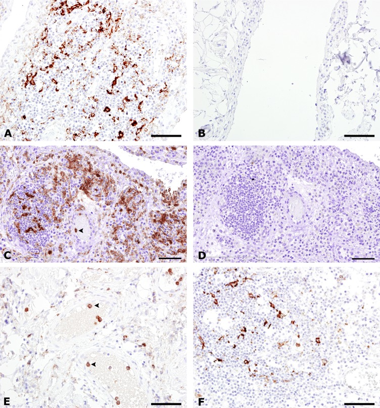
Fig 1. C5aR expression in the synovium from rheumatoid arthritis, osteoarthritis psoriatic arthritis, and non-inflammatory control.
Immunohistochemical staining for C5aR (A, B, C, E and F), and for the IgG2a isotype control antibody (D). Synovium from patients with RA undergoing joint replacement is shown in (A), and synovectomy, (C, D). Synovium from patients with OA is shown in (E), and PsA in (F). Synovium from a non-inflammatory control is seen in (B). Numerous infiltrating C5aR+ cells (in brown) are seen in A, C and F. Intravascular C5aR+ neutrophils are seen in C and E (arrowheads). Note the lack of immunoreactivity in the non-inflammatory control (B). Nuclei (in blue) were counterstained with haematoxylin. Bars: (A, B, E and F) 50 μm, (C and D) 100 μm.

