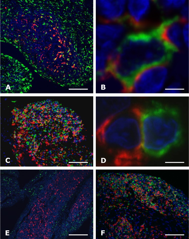
Fig 2. C5aR expression by macrophages and neutrophils, but not by T cells in the synovium of rheumatoid arthritis.
Double immunofluorescence staining for C5aR (red signal A—F) and either CD68+ macrophages (green signal A, B and C), CD3+ T cells (green signal in D), or MPO+ neutrophils (green signal in E and F) of the synovium from rheumatoid arthritis patients undergoing joint replacement (A, B, D and E) or synovectomy (C and F), analysed by confocal microscopy (A, B, D and E) and epifluorescence microscopy (C and F). Framed area in E is seen in B. Note the co-localization of C5aR and CD68+ macrophages in A, B and C, and MPO+ neutrophils F, and lack of co-localization with a CD3+ T cell in D. The nuclei (in blue) were counterstained with Hoechst. Bars: (A) 100 μm, (B and D) 5 μm, (E) 500 μm, (C and F) 50 μm.

